Introduction to the Circulatory System
The Circulatory system in most animals, including humans, utilizes blood, a mixture of water, interstitial fluid, plasma, platelets, and other cells like leukocytes (White Blood Cells)! The purpose of blood is to transport specific nutrients such as oxygen and other molecules, like glucose from the digestive system, to each individual cell in an organism’s body, and waste, such as carbon dioxide (CO2) and urea out of the body via the lungs or kidneys.
In small, simple animals, this is easily done in what is called an Open Circulatory System, usually present in invertebrates like insects, arthropods, and mollusks. An open circulatory system is absent of any blood vessels, however does have a blood pump (heart), but has no separation of blood or interstitial fluid.
Oppositely, a Closed Circulatory System is found in a few invertebrates like earthworms, squids, and octopuses, but is in nearly all vertebrates like humans. The structure of a closed circulatory system is a large, complex network of tubes called blood vessels which act as transports for the blood to travel throughout the body. The blood never exits the vessels (unless in the case of a wound) and is pushed via high pressure from a pump, commonly referred to as the heart. The more complex and active the animal, the larger and more complex the heart must be. Please note that just because an animal has a more complex heart, with more chambers, it is not always a larger animal than an organism with a simpler heart. (For your benefit, it is good to know that Red Blood Cells can also be referred to as Erythrocytes.)
Grasshopper (Open Circulatory System) Versus Earthworm (Closed Circulatory System)
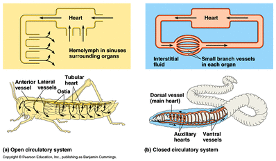
Grasshoppers are the best example of an open circulatory system to give on exams and earthworms are very simple animals that show exactly what a closed circulatory system is like. Disregard the formal names of the structures in this diagram (anterior vessel, auxiliary hearts, ostia, etc.) however, interstitial fluid and hemolymph are necessary terms to remember. Hemolymph is the open circulatory system equivalent to a close circulatory system's "blood." Interstitial fluid is the fluid found in both circulatory systems that provides liquid to the entire body for nutrients and wastes to cross via Simple Diffusion (Simple Diffusion requires a moist membrane to move materials).
How does Blood carry Oxygen?
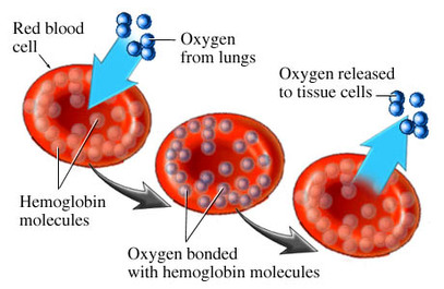
Blood has the ability to carry oxygen with help of a famous and very helpful protein called hemoglobin. Hemoglobin is found all over any healthy red blood cell and contains iron which is used as the binding site for oxygen molecules since they attract O2 molecules. Why oxygen MUST be transported in this manner is that oxygen is not readily soluble in water (major component of blood, part of plasma), unlike carbon dioxide which can be dissolved into carbonic acid in the blood, AND can be carried using a hemoglobin molecule (different binding site however). When pH becomes too low (due a large amount of carbonic acid), hemoglobin binds to oxygen with less affinity and releases it where CO2 is high. Then once the oxygen has been given to the cells, the blood carries the carbon dioxide to the lungs for excretion and with a rise in pH (due to a lack of carbonic acid) hemoglobin has more affinity for O2 and can receive more oxygen to transport across the body once more. This process is known as the Bohr Shift however is a more advanced concept that does not need to be memorized yet for the Circulatory System.
Varied Chambered Hearts
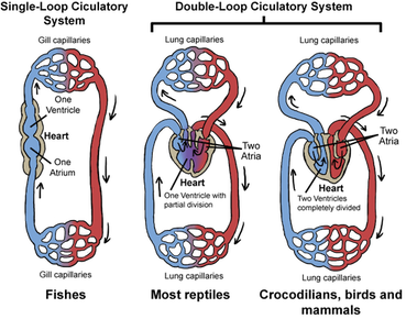
Hearts are usually seen to be either 2, 3, or 4 chambered, meaning areas of the heart where blood is stored and pumped throughout the organism. Typically fish have 2 chambered hearts which does grant them a heart to pump blood to send necessary nutrients into the body's cells and wastes out of the body, but has a weakness of low pressure. 3 Chambered hearts on the other hand (found is amphibians and reptiles) provide higher pressure but is unable to transport a great amount of O2 into the body as deoxygenated blood and oxygenated blood mix within the heart. Finally, 4 chambered hearts (found in large, active animals like mammals) not only provide high pressure for effective transport of nutrients, but separates deoxygenated blood from oxygenated blood. Also, usually animals with 4 chambered hearts are endothermic or "warm-blooded" and thus require ten times as much oxygen as ectotherms or "cold-blooded." A 4 chambered heart is the most efficient and powerful tool in a circulatory system and is what will be studied on this webpage.
However, from an evolutionary perspective, why don't all animals have a 4 chambered heart since it is obviously the best choice? The answer is a 4 chambered heart costs a high amount of energy to operate so animals which eat far less than mammals or are unable to get as many nutrients., cannot afford a 4 chambered heart. Usually it's because many animals like a snake who only digest a meal about once a month does not need a more complex heart than 3.
Fun fact: Birds can also have 4 chambered hearts similar to mammals but are not closely related. This is an example of Convergent Evolution which you will learn in Miss Rossman's class.
The Pathways of the Circulatory System
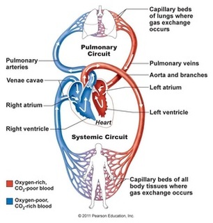
The diagram to the left shows how blood travels throughout the body in an easy to understand illustration. Let's just begin the in the Left Atrium (LA) located at the top of the heart on THE ORGANISM'S left side, NOT YOURS. Oxygenated blood that just came from the lungs and sent through the Pulmonary Vein enters the Left Atrium and is pumped through the heart into the Left Ventricle or bottom left of the heart. To get into the left ventricle from the left atrium, the blood had to pass through the atrioventricular valve (please note both the mitral valve and tricuspid valve, as seen in the diagram below, can be referred to both as atrioventricular valves for simplicity) so when blood is pumped again, the valve halts the blood from going in the wrong direction as seen in the next picture. Then once the blood passes the atrioventricular valve and is in the left ventricle, the blood passes through the semi-lunar valve or aortic valve, and into the Aorta (the main artery of the heat) which is shown as the red blood vessel that moves blood away from the heart. (artery) Then the nutrients from the blood are exchanged via diffusion (explained later on), to all of the body's cells in capillaries which are incredibly small and thin blood vessels that provide a high amount of surface area for efficient exchange of nutrients and waste. Then once deoxygenated blood exits the capillaries and enters the veins (blood vessels that send blood TOWARD the heart, NOT vessels that contain deoxygenated blood) they return to the heart via either the posterior vena cava (if blood came from the legs) or superior vena cava (if blood came from the brain) and into the Right Atrium (RA). Then the blood goes through the next atrioventricular valve and into the Right Ventricle (RV). Then, the blood is pumped through the next semi-lunar valve, or pulmonary valve, into the Pulmonary Artery (carries unoxygenated blood AWAY from the heart) and into the lungs where carbon dioxide is excreted and oxygen is given to the red blood cells to transport to all the other cells in the body. Then the blood enters the Pulmonary Vein (carries oxygenated blood TOWARD the heart) and into the Left Atrium of the heart, restarting the Cardiac Cycle.
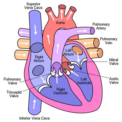
This is a greatly detailed diagram so please study it well. Please note that the Right Atrium/Right Ventricule pumps oxygen poor blood into the pulmonary artery (light purple vessel shown here) which goes to either the left or right lung, to become oxygenated, and then goes through the Pulmonary vein to enter the Left Atrium of the Heart.
|
|
Guiding Questions1. What is Simple Diffusion and how does it work?
2. What are the pathways of the blood? 3. What does the Pulmonary Artery carry (deoxygenated or oxygenated blood)? 4. What does the Pulmonary Vein carry (deoxygenated or oxygenated blood)? 5. Why do mammals have 4 chambered hearts? |
The above video addresses both the circulatory system and respiratory system, but is still a great video to look at for our circulatory system guide. Both videos here are required to be watched.
|
|
Guiding Questions1. How Does a 2, 3, and 4 chambered heart work respectively?
2. What color is unoxygenated blood? (hint: NOT BLUE) 3. What stops blood from going backwards in veins where blood pressure is low? 4. What is Systole and what is Diastole? Which chambers of the heart control which? 5. What is "Lub" and "Dub?" (Yes, these are their scientific names). 6. What is the Sinoatrial Node? |
Artery/Vein Structure and Why?
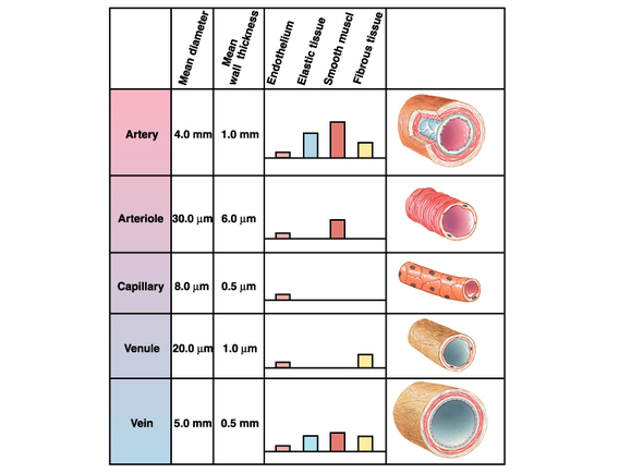
Arteries are the vessels that carry blood away from the heart and experience the highest pressure from it. This pressure must be able to reach the entire body without much freedom to fail so the pressure is of course relatively intense. To adapt to this, arteries have very large, flexible, and strong walls to contain blood without bursting from the pressure. Arterioles are structures that branch off from the large arteries near the heart to reach the capillaries. The capillaries are thin (more thin than the diameter of a red blood cell) and fragile meaning that if pressure were as high here as at the actual heart, the capillaries would explode so they are located far away from the pressure to perform their job without bursting. Remember from earlier that capillaries are immensely thin to provide plenty of surface area for diffusion (as you will, or already have learned is the same reason for having a huge number of alveoli in the respiratory system). Venules are the equivalent of arterioles for veins as they are increasingly large (from small diameter) vessels to maintain the pressure from the capillaries. Finally, once the blood reaches a vein, there is a small problem as the pressure from the heart has decreased dramatically and pooling of blood occurs in the veins, which is why they are large in diameter and elastic. So how does this blood get back to the heart? Muscle contractions from moving your arms or legs squeeze your enlarged veins and push blood back to the venae cavae and into the heart to renew their pressure. The Heart is not the only thing in your body that pump blood (which is why physical activity is important for healthy blood circulation)!
Circulatory System's Role in Homeostasis?
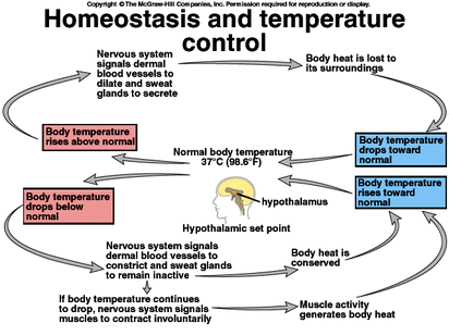
Homeostasis is the process by the endocrine system to regulate internal body conditions to be constant and regulated. For example, if our body temperature gets too low, less activity occurs in our body, lowering survival rate and if it gets lower than 68 degrees Fahrenheit, the heart (in humans) stops beating entirely, resulting in death. If temperature gets too high, then proteins become denatured (misshaped) and since "structure determines function" as Miss Rossman will tell you many times for proteins, the proteins become useless and again, we die.
Blood plays a monumentally crucial role in homeostasis (as the endocrine system guide should tell you) as it sends specific hormones all over the body to communicate messages that the nervous system is unable to accomplish. Blood and the circulatory system is both used for communication and homeostasis (example: blood glucose levels and temperature).
How blood can regulate temperature is, when signaled to keep the body warm, blood vessels constrict (to keep inner organs warm and not outside skin) and use the warm energy they receive from the digestive system to keep the body at a perfect 97 to 99 degrees Fahrenheit. When signaled to cool off the body (along with sweat) blood vessels dilate (get bigger to have more contact with the cooler, outside environment) to reduce body temperature.
"Lub" and "Dub"
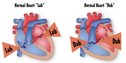
The famous and iconic, rhythmic sounds of the heart known as "lub" and "dub" are essentially just the sounds of valves in the heart closing.
"Lub" (which comes first) is the sound of blood recoiling against the atrioventricular valves in the heart between the left/right atrium and left/right ventricle.
"Dub" (which comes next) is the sound of blood recoiling against the two semilunar valves between the right ventricle and pulmonary artery and left ventricle and aorta.
When blood does squirt through defected, closed valves, the following hissing sound is what is known as a Heart Murmur.
Sinoatrial Node
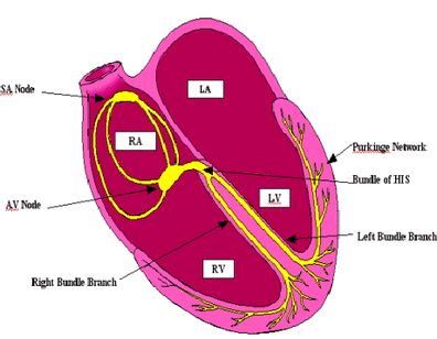
The Sinoatrial Node (denoted as SA in the picture) is the pacemaker of the heart which is responsible for the involuntary, rhythmic beatings of the heart in our everyday lives. It is basically a master nerve whose only purpose is to constantly communicate with the cardiac muscles of the heart to keep it beating at varying rates (depending on our situation) throughout our lives.
When in danger, the sinoatrial node will command the heart to beat faster to facilitate respiration and allow the body to initiate the "fight or flight" response with an increase of energy to expend. When sleeping or resting, the sinoatrial node will command the heart to beat slower, which will conserve energy for later use.
When in danger, the sinoatrial node will command the heart to beat faster to facilitate respiration and allow the body to initiate the "fight or flight" response with an increase of energy to expend. When sleeping or resting, the sinoatrial node will command the heart to beat slower, which will conserve energy for later use.
Systole and Diastole (Blood Pressure)
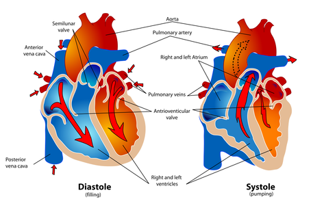
While conventional "heart beats per minute" may give one reading of blood pressure, the concepts of systole and diastole display the whole picture. Systolic pressure is the pressure at which blood is pumped out of the heart by the ventricles and Diastolic pressure is the pressure at which the blood fills the atriums and ventricles of the heart.
When providing a measurement of blood pressure, it is always represented as Systole/Diastole where systole is the peak pressure and diastole is the minimum pressure. A normal blood pressure is 110/70 where 110 is systolic pressure (naturally higher in value) and 70 is diastolic pressure (naturally lower in value).
High blood pressure is known as Hypertension and is typically a value of 150/90 where 150 is Systolic pressure and 90 is Diastolic pressure.
The Lymphatic System
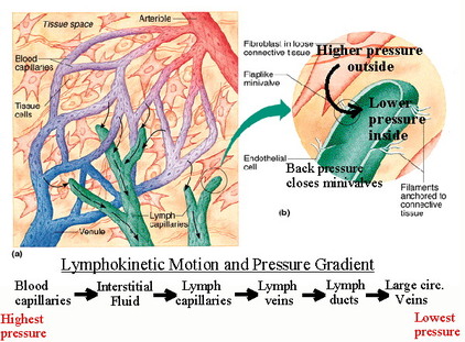
The Lymphatic System is a series of vessels that is parallel to the Circulatory System's blood vessels. The lymphatic system's lymph vessels are intertwined amongst the blood vessels are responsible for the collection of interstitial fluid, maintaining blood volume and protein concentration, and transportation of White Blood Cells (WBCs for short or A.K.A. Leukocytes). The lymphatic system defends against infection and makes certain that the blood vessels transport blood to transport nutrients efficiently.
The picture to the left is more advanced than what is required for you to know but what is important to know from it is that blood vessels are not the only tubes that transport liquid throughout the body and that there is a system in the body for controlling interstitial fluid in blood.
There is easy transport of interstitial fluid and WBCs via diffusion (very little distance since both blood and lymph vessels are in near direct contact with one another) and if blood is in need of materials from the lymphatic system, interstitial fluid and WBCs are drained in Superior/Posterior Venae Cavae and the Right Atrium.
Bonus Video!To give an even higher understanding of the Circulatory System and the life of an average erythrocyte (Red Blood Cell) and the Bohr Shift. We recommend watching after this page has been read fully or not at all if you think you're fine. Good luck on your quiz!
Again, the Bohr Shift will NOT appear on your quiz, but is a great concept to understand for later on in the year. |
|
Answers to Guiding Questions
Crash Course
1. Simple Diffusion is the passive movement (no energy input from the cell) of molecules through a moist membrane. It must be moist in order for the molecules to pass through the membrane.
2. Left Atrium -> atrioventricular valve -> Left Ventricle -> semilunar valve or Aortic valve -> Aorta -> either toward the head or to the legs via arteries -> capillaries -> veins -> posterior/inferior vena cava if coming from the legs...superior vena cava if coming from the head -> Right Atrium -> atrioventricular valve -> Right Ventricle -> semilunar valve or Pulmonary valve -> Pulmonary Artery -> right or left lung -> Pulmonary Vein -> Left Atrium
3. Pulmonary Artery carries deoxygenated blood
4. Pulmonary Vein carries oxygenated blood
5. Mammals have 4 chambered hearts because they are far more active than reptiles and amphibians, so they require more oxygen. Mammals also typically live on land and require a heart that can grant high pressure to make sure that blood can get around the whole body despite the downward effect of gravity. 2 Chambered hearts cannot provide this pressure and do not need to since they live in water where gravity's effects are negligible. 3 Chambered hearts cannot grant mammals the amount of oxygen they need to stay so active.
Bozeman
1. 2 chambered hearts have one ventricle and one atrium and pumped blood through their body with gills being the equivalent of lungs to present blood with oxygen. 3 chambered hearts have 2 atriums and one ventricle which has high pressure but mixes deoxygenate blood with oxygenated blood so it can't bring a great mount of oxygen to body. 4 chambered hearts have 2 atrium and 2 ventricle which separate deoxygenated and oxygenated blood, and provide high pressure to the body.
2. Blood is always red due to the iron in the hemoglobin of the red blood cells. The myth that deoxygenated blood is blue is complete and utter bunk. Veins may appear blue under our skin but I can assure you they carry red blood.
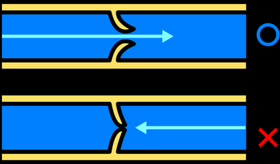
3. What halts blood from falling in the opposite direction are numerous valves in the veins. Once blood enters through the valves, they cannot exit the same way. This allows blood to pool in veins and help them reach the right atrium to renew their pumping more efficiently.
4. Systole is the pumping of blood by the right and left ventricle into the aorta (by left ventricle) and pulmonary artery (right ventricle), and diastole is the filling of blood into the right/left atrium and right/left ventricle. Please note heart pressure is given by Systole/Diastole.
5. "Lub" is the sound of the closing of the two atrioventricular valves in the heart and "dub" is the sound of the closing of the two semilunar valves in the heart or Aortic and Pulmonary Valve.
6. The Sinoatrial Node is merely the bundle of nerves that control the pace the heart beats at and to make sure it beats continuously throughout our whole life.
Components of Blood/Blood Conditions Online Document
1. Red Blood Cells or RBCs (erythrocytes)are cells void of any nucleus or organelles and only contains hemoglobin to transport oxygen throughout the body.
2. White Blood Cells or WBCs (leukocytes) fight infection for the body by destroying pathogens.
3. Platelets are not actually true cells and do not have a nucleus. They are fragments of cells which are only used to clot open wounds in the body to prevent massive loss of blood. When platelets cannot do this, it is a condition called hemophilia.
1. Simple Diffusion is the passive movement (no energy input from the cell) of molecules through a moist membrane. It must be moist in order for the molecules to pass through the membrane.
2. Left Atrium -> atrioventricular valve -> Left Ventricle -> semilunar valve or Aortic valve -> Aorta -> either toward the head or to the legs via arteries -> capillaries -> veins -> posterior/inferior vena cava if coming from the legs...superior vena cava if coming from the head -> Right Atrium -> atrioventricular valve -> Right Ventricle -> semilunar valve or Pulmonary valve -> Pulmonary Artery -> right or left lung -> Pulmonary Vein -> Left Atrium
3. Pulmonary Artery carries deoxygenated blood
4. Pulmonary Vein carries oxygenated blood
5. Mammals have 4 chambered hearts because they are far more active than reptiles and amphibians, so they require more oxygen. Mammals also typically live on land and require a heart that can grant high pressure to make sure that blood can get around the whole body despite the downward effect of gravity. 2 Chambered hearts cannot provide this pressure and do not need to since they live in water where gravity's effects are negligible. 3 Chambered hearts cannot grant mammals the amount of oxygen they need to stay so active.
Bozeman
1. 2 chambered hearts have one ventricle and one atrium and pumped blood through their body with gills being the equivalent of lungs to present blood with oxygen. 3 chambered hearts have 2 atriums and one ventricle which has high pressure but mixes deoxygenate blood with oxygenated blood so it can't bring a great mount of oxygen to body. 4 chambered hearts have 2 atrium and 2 ventricle which separate deoxygenated and oxygenated blood, and provide high pressure to the body.
2. Blood is always red due to the iron in the hemoglobin of the red blood cells. The myth that deoxygenated blood is blue is complete and utter bunk. Veins may appear blue under our skin but I can assure you they carry red blood.
3. What halts blood from falling in the opposite direction are numerous valves in the veins. Once blood enters through the valves, they cannot exit the same way. This allows blood to pool in veins and help them reach the right atrium to renew their pumping more efficiently.
4. Systole is the pumping of blood by the right and left ventricle into the aorta (by left ventricle) and pulmonary artery (right ventricle), and diastole is the filling of blood into the right/left atrium and right/left ventricle. Please note heart pressure is given by Systole/Diastole.
5. "Lub" is the sound of the closing of the two atrioventricular valves in the heart and "dub" is the sound of the closing of the two semilunar valves in the heart or Aortic and Pulmonary Valve.
6. The Sinoatrial Node is merely the bundle of nerves that control the pace the heart beats at and to make sure it beats continuously throughout our whole life.
Components of Blood/Blood Conditions Online Document
1. Red Blood Cells or RBCs (erythrocytes)are cells void of any nucleus or organelles and only contains hemoglobin to transport oxygen throughout the body.
2. White Blood Cells or WBCs (leukocytes) fight infection for the body by destroying pathogens.
3. Platelets are not actually true cells and do not have a nucleus. They are fragments of cells which are only used to clot open wounds in the body to prevent massive loss of blood. When platelets cannot do this, it is a condition called hemophilia.
The Circulatory System Created By Samuel and Daniel Mueller
We Hope you found this page helpful and torturous
P.S. We take full credit for the surplus of Crash Course videos :)
P.S. We take full credit for the surplus of Crash Course videos :)
